Anterior Cervical Discectomy and Fusion
Anterior Cervical Discectomy and Fusion is a surgical procedure to remove a herniated disc in the neck, where an incision is made in the throat area to access and remove the disc, and a graft is inserted to fuse the bones together above and below the disc, ACDF surgery may be an option if treatment fails Natural or medicines relieve neck or arm pain caused by compressed nerves, and patients are offered a service in installments for operations at operazi.
Information about Anterior Cervical Discectomy and Fusion
● Discectomy literally means 'disc cutting'. A discectomy can be performed anywhere along the spine from the neck (cervical) to the lower back (lower back).
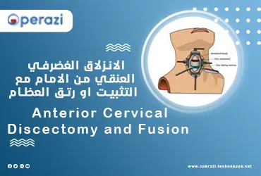
● The surgeon accesses the damaged disc from the front (anterior) part of the spine through the throat area and by moving the muscles of the neck, trachea and esophagus aside, thus exposing the disc and bony vertebrae.
● The surgery is more accessible from the front of the neck than from the back (back) because the disc can be accessed without disturbing the spinal cord, spinal nerves, and strong neck muscles.
● Depending on your symptoms, one (single-level) or more (multi-level) discs may be removed.
● After the disc is removed, the space between the bony vertebrae becomes empty, and to prevent the vertebrae from collapsing and rubbing together, a spacer bone graft is inserted to fill the open disc space.
● The graft acts as a bridge between the two vertebrae to create a spinal fusion.
● The graft and vertebrae are held in place with metal plates and screws.
● After surgery, the body begins its natural healing process and new bone cells grow around the graft.
● After 3 to 6 months, bone grafts should join the two vertebrae and form one solid piece of bone. The organs and fusions work together, similar to reinforced concrete.
● After the fusion, you may notice some loss of movement, but this varies according to the movement of the neck before surgery and the number of levels fused.
● If only one plane is incorporated, you may have similar or better range of motion than you had before surgery.
● If more than two levels are combined, you may notice limits in turning your head and looking up and down.
● Artificial disc replacements that preserve motion have emerged as an alternative to fusion. Similar to a knee replacement, the artificial disc is inserted into the damaged joint space and preserves movement, while fusion eliminates movement.
See also: total proctocolectomy with or without colostomy
What are the sources of bone grafts?
Bone grafts come from several sources, and each type has advantages and disadvantages:
1- Autograft bones come from you:
● The surgeon takes bone cells from your hip (iliac crest).
● This graft has a higher fusion rate because it contains cells and proteins that grow in the bones.
● The disadvantage is the pain in the femur after surgery.
● A bone graft is taken from the hip at the same time as the spine surgery, the thickness of the cut bone is about half an inch - not the entire thickness of the bone is removed, only the top half layer.
2- Allograft bone comes from a donor (cadaver):
● Bones are collected from the bone bank of people who have agreed to donate their organs after their death.
● This graft does not contain cells or proteins that grow in the bone, however it is readily available and eliminates the need to harvest bone from the hip.
● The graft is shaped like a round cake and the center is filled with a crust of live bone tissue taken from your spine during surgery.
3- Plastic or ceramic bait:
● Or man-made absorbable compounds.
● Often called cages, this graft material is filled with a shell of live bone tissue taken from your spine during surgery.
candidate for Anterior Cervical Discectomy and Fusion
You may be a candidate for a discectomy if you have:
● Diagnostic tests (MRI, CT scan, myelography) show that you have a herniated disc or a degenerative disc.
● Significant weakness in your hand or arm.
● Arm pain is worse than neck pain.
ACDF may be useful in treating the following conditions:
● Disc bulge and herniation: The gelatinous substance inside the disc can bulge or rupture through a weak area in the surrounding wall (annulus). Irritation and swelling occur when this material compresses and puts painful pressure on the nerve.
● Degenerative disc disease: When discs wear out normally, bone spurs form and face joints inflame. Discs dry out, shrink and lose their elasticity and cushioning properties. Disc spaces become smaller.
What happens during the process of Anterior Cervical Discectomy and Fusion?
There are seven steps to this procedure, the process generally takes 1 to 3 hours.
Step one: prepare the patient
You will sleep on your back and you will be sedated. Once you sleep, your neck area is cleaned and groomed. If a fusion is planned and your bone will be used, the hip area will also be prepared for a bone graft. If a donor bone is used, a hip incision is not necessary.
Step 2: Make an incision an incision is made
The surgeon creates a tunnel into your spine by moving your neck muscles aside and pulling your windpipe, esophagus and arteries. Finally, the muscles that support the front of the spine are lifted and set aside so that the surgeon can clearly see the bony vertebrae and discs.
Step 3: Locate the damaged disk
With the help of a fluoroscope (a special X-ray), the surgeon passes a thin needle into the disc to locate the affected vertebra and disc. The bones of the vertebrae are spread above and below the damaged disc by means of a special connective.
Step 4: Remove the disc
it is cut off the outer house of the disk. The surgeon removes about two-thirds of the disc using small grasping tools, then looks through a surgical microscope to remove the rest of the disc. The ligament that passes behind the vertebrae to access the spinal canal is removed.
Step 5: Decompress the nerve
Bone spurs that press on the nerve root and enlarges the foramen, through which the spinal nerve exits, are removed with a drill. This procedure, called a puncture hole, gives your nerves more room to exit the spinal canal.
See also: Excision of tumor or cyst from liver / pancreas
Step 6. Preparation of the osseointegration
With a drill, the open disc space at the top and bottom is prepared by removing the outer cortical layer of bone to expose the blood-rich spongy bone inside. This space will contain the bone graft material that you and your surgeon have chosen:
● Femur graft: An incision is made in the skin and muscle above the top of the femur. Next, a chisel is used to cut the hard outer layer (cortical bone) to the inner layer (spongy bone). The inner layer contains the cells and proteins that grow bone. Then the bone graft is formed and placed in the space between the vertebrae.
● Bone Bank: A cadaver's bone grafts or bioplastic cage are filled with leftover bone sawdust that contains cells and proteins that grow in the bones. Then the bait is inserted into the hole space.
The bone graft is often reinforced with a metal plate that is attached to the vertebrae to provide stability. During fusion, X-rays are taken to check the position of the graft, plate, and screws.
.


00 التعليقات