image description

Operazi care About Your Life & health,Operazi A platform For surgeries with Financial installment


Operazi is the first intelligent platform facilitating global access to surgical and medical interventions providers, allowing easy search, simple browsing, comparing and booking surgical operations and other medical procedures. Along several other services as electronic payment, payments by installments, post operative care, medical tourism services, and insurance facilitation services. All at a click of a button!

A Complete Medical Surgery Portal

Operazi is the first platform For surgeries with Financial installment In Egypt. Our comprehended health system offers you convenient clinics and hospitals to Provide preventative and follow-up care.

Operations Offers

No Data To Display It

Valuable Feedback

Mohamed Kamal Ismail Dowidar

Mohamed Kamal Ismail Dowidar

Latest Articles

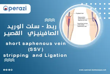
short saphenous vein (SSV) stripping and Ligation is a surgical procedure we use to remove severely damaged veins in the legs, which are causing venous insufficiency or varicose veins. Once the damaged vein is removed, it will take over other veins in your body to allow blood to flow in that part of your leg. Our Cardiovascular Services at operazi offer the latest innovations in vascular care for patients. operazi can perform the short saphenous vein (SSV) stripping and Ligation in installments. For our services, click here.

Who is a Candidate for short saphenous vein (SSV) stripping and Ligation?

You good candidate if you:

• They have severe venous insufficiency.

• They have very large varicose veins.

• Are at risk of developing venous ulcers or already have skin ulcers.

• You experience frequent pain or heaviness in your legs.

• You have severe leg pain.

• Damaged veins in places where superficial veins (closest to the surface of the skin) connect to deep veins.

A vascular surgeon may discuss other options instead of surgery if you:

• Older.

• You have other medical conditions that put you at greater risk of complications from the surgery.

• You have poor blood circulation in the arteries in your leg (the large blood vessels that carry oxygen from your heart to your body).

• You have swelling or fluid buildup in the area of ​​the varicose veins due to lymphedema.

• You have a skin infection.

• They are more prone to blood clots.

•pregnant.

We at operazi offer both surgical and non-surgical treatment options to help you customize the treatment plan that best suits your needs.

See also: surgical prostatectomy

What do you expect on your first date with operazi?

At your first appointment, you'll meet with a vascular surgeon who will ask about your medical history and discuss your concerns about venous insufficiency and/or varicose veins. They will examine your legs and look for other symptoms, such as swelling, discoloration of the skin or varicose veins. Any invisible symptoms, such as itching or pain, should also be discussed with your provider.

We may order an ultrasound to see what's going on inside your veins. We'll run a small hand-held device (called a transducer) over the top of your skin to see pictures of the veins inside your legs. These ultrasound images will help us create the best treatment plan for you, also you can search for a vascular surgeon in operazi

How do you prepare for a short saphenous vein (SSV) stripping and Ligation surgery?

Vein ligation and stripping is a minor surgery. You will sleep under general anesthesia. It is important to follow your surgeon's instructions to prepare for surgery.

• You will not be able to drive on your own after you are out of anaesthesia.

• Your surgeon and anesthesiologist will meet with you before surgery to discuss the procedure and answer any questions you have.

What happens during a short saphenous vein (SSV) stripping and Ligation surgery?

• The vascular surgeon will make two small incisions (incisions) - one in the groin area (near the top of the damaged vein) and one in the thigh or calf (below the vein).

• The surgeon will tie off the upper part of the vein to stop the blood flow. A thin, flexible device will be passed through the damaged vein. The surgeon will use the device to pull the entire vein out through an incision (cut) below the vein.

Once the vein is removed, the surgeon will stitch up your incisions and place dressings over them. This procedure is called phlebectomy or microdissection and can be done at the same time as vein stripping and ligation.

After surgery is complete, your surgeon will place gauze and an ACE-compression roll on your treated leg. You will be taken to the recovery room to rest and wake up from the anesthesia. You'll be in the recovery room for one to two hours before a family member or friend can take you home. The surgeon will give you compression stockings to wear the next day after the dressings are taken off, and our vascular surgeons in operazi can treat all types of conditions that lead to venous insufficiency and varicose veins.

Complications of ligation of the short saphenous vein

The most common risks include:

• Infection after surgery.

• Bleeding.

• Blood clots.

• Scarring or bruising at the site of surgery.

• Injury to the nerves around the vein.

• reaction to anesthesia.

Recovery time with short saphenous vein (SSV) stripping and Ligation surgery

You will need to wear compression stockings for about two to four weeks after surgery for your leg to recover. Healing time may vary depending on the number and location of the veins stripped. Your surgeon will discuss the healing process with you in more detail.

Signs of recovery after surgery

• You may feel some slight pain after the vein ligation and stripping surgery. If necessary, you can take over-the-counter pain relievers to control the pain. Your pain will improve as your legs heal.

• Most people experience significant bruising immediately after surgery. Postoperative bruising takes up to three weeks to heal.

• It will take about three weeks for your incisions to heal and the stitches to dissolve.

Bathing and showering after surgery

• Don't shower until your doctor tells you can take off your bandages and compression stockings, usually the day after surgery.

See also: drainage of renal abcess

Resume daily activities

Most people will be able to get around within a few hours after surgery. As your legs recover over the next two to four weeks, continue to elevate your legs as much as you can while sitting.

It's important to move your legs and keep blood flowing as much as possible once your surgeon has authorized you to resume normal activity. We suggest you:

• Walk around once every hour for 10 to 15 minutes at a time.

• Flex and direct your feet while sitting or lying down.

• Increase your intake of exercise each day as your legs begin to feel better and the cracks (wounds) heal.

Do not do any strenuous exercise such as lifting heavy objects until your surgeon tells you to do so safely.

Driving and back to work

You will not be able to drive for at least three days. Talk to your doctor about when you can resume driving. Most people can return to work one week after recovery. If your job involves physical stress, standing for long periods of time, or other activities that can put pressure on your legs, your doctor may tell you to wait longer before returning to work.

;

renal /Peripheral arteries angioplasty is a technique to open narrowed blood vessels in the kidneys. A very small balloon attached to a thin tube (catheter) is inserted into a blood vessel through a small incision in the skin. The catheter is inserted under X-ray guidance to the site of the narrowed artery. The balloon is inflated to open the artery. Sometimes a small metal scaffold called a stent is also inserted to keep the blood vessels open. Catheters and stents often replace graft surgery in the treatment of arterial problems, and blockage of the renal artery can lead to serious problems, and it is wise to seek medical attention without delay. operazi can perform the renal /Peripheral arteries angioplasty in installments. For our services, click here.

What is the process of renal /Peripheral arteries angioplasty by balloon?

Your interventional cardiologist or vascular specialist may recommend renal /Peripheral arteries angioplasty to open blocked arteries and restore blood flow. renal /Peripheral arteries angioplasty is a procedure that is performed in the catheterization laboratory of the hospital. You will lie on a table and be lightly sedated to help you relax, but you will remain awake throughout the procedure.

The renal artery is the main blood vessel of the kidneys. Renal artery angioplasty and stenting is done to treat renal artery stenosis, also called renal artery stenosis. Using an X-ray as a guide, a small plastic tube is placed into the narrowed artery. A special balloon is inflated onto the tube to open the narrowed part of the artery. An expandable tube called a stent is then placed to keep the artery open.

See also: surgical prostatectomy

What are the steps of the process of renal /Peripheral arteries angioplasty by balloon?

You will be asked to lie on your back on an X-ray bed. Staff will clean the skin on your thigh or arm and place sterile drapes over you. Then they inject a local anesthetic, which may sting a little.

1- Renal catheterization

• A thin flexible tube called a catheter is inserted into the artery and passed through the arteries to the blockage. (Because there are no nerves in the arteries, you won't feel the catheter.) An X-ray camera and images of the arteries (shown on a TV screen in the catheter lab) will help the doctor guide the catheter into the blockage.

• The dye is injected through the tube. The tube is X-rayed to make sure it is in the right place.

• A thin, thin wire will then be passed through the narrowed part of the artery. It serves as support to place the small balloon through the blockage.

• Next, a balloon catheter - a long, thin, flexible tube with a small, uninflated balloon at its tip - will be inserted into the artery where the artery is narrowed.

• Once in position, the balloon will be inflated to open the artery.

2-mode stent

In most cases, a small metal mesh tube called a stent is placed.

• It will be placed to keep the artery open and reduce the risk of narrowing the artery again.

• After the stent is placed, your doctor will remove all catheters.

And connect with operazi, the best and first installment site for vascular operations.

What happens after the renal /Peripheral arteries angioplasty?

• Most renal artery patients treated with angioplasty and stenting are discharged from the hospital 12 to 24 hours after catheter removal.

• Many patients are able to return to work within a few days up to a week after the operation.

• If the intervention involves inserting a stent, your cardiologist or vascular specialist will prescribe blood-thinning medications, such as aspirin and clopidogrel, usually for a month to a year.

• The dye will leave your body in the urine. You won't notice it because it's colorless, so drink plenty of fluids to help get rid of the dye.

• It is very important that you follow your doctor's instructions and take your medicines as prescribed by your doctor. Even after you begin to feel better, you should never stop taking your medications at any time without talking with your interventional cardiologist.

When do you call your doctor?

After any procedure, it's important to stay in touch with your doctor. After the balloon renal /Peripheral arteries angioplasty, your health care provider will recommend that you monitor the area where the catheter was inserted. If the wound increases in size, turns red, empties a thick yellow/brown substance, or is painful, even when there is no pressure, contact your doctor. A painful lump may be the biggest sign that the puncture hole is not healing properly or is leaking blood.

Other reasons to call your doctor include:

• If you have a fever.

• If you have swelling - with or without pain - anywhere in the leg where the catheter was inserted.

And contact the operazi website, the best site that offers a medical consultation booking service.

See also: drainage of renal abcess

When do normal activities resume after your procedure?

Always use your direct doctor's instructions about the right level of activity for you. Once you leave the hospital, your post-operative care will be twofold.

• You will need to take care of yourself by taking the medication exactly as prescribed, as well as making lifestyle changes, such as exercising, improving your diet and, if you are a smoker, quitting.

• You should also have at least one follow-up appointment with your interventional cardiologist or other qualified specialist if you are not able to return to your physician. At this appointment, the interventional cardiologist will check the catheter insertion site to make sure it is healing properly.

;

The aorta is a large artery that carries oxygen-rich blood from the left heart chamber to the rest of the body. A rupture in the small blood vessels that supply the walls of the aorta leads to an intra-aortic hematoma. Surgical treatment of an aortic IMH is performed as a result of aortic dissection because there is bleeding in the aortic wall, usually from the vessels without internal rupture, and it forms part of the acute aortic syndrome along with Combined with penetrating atherosclerotic ulcers and classic aortic dissection. Usually, intra-aortic hematomas appear in elderly hypertensive patients, and the same condition may also develop as a result of acute chest injury, aortic wall injury or penetrating atherosclerotic ulcer. operazi can perform the Surgical treatment of an aortic IMH in installments. For our services, click here.

What is Surgical treatment of an aortic IMH?

Surgical treatment of an aortic IMH is performed when an intra-aortic hematoma occurs due to damage to the tiny blood vessels along the aortic wall. Blood leaks from the blood vessels and accumulates within the walls. It is also considered a form of aortic dissection (caused by a tear in the inner layer of the aorta).

 What causes aortic IMH?

The main factor that leads to an aortic IMH is unknown. However, there are certain factors such as:

• Leaking blood from the middle layer of the aorta leads to the formation of blood-filled spaces, which weakens the wall of the aorta without tearing the outer layers of the aorta.

• High blood pressure causes arterial walls to thicken, affecting normal blood flow.

• Atherosclerosis causes the buildup of fats (plaques) and other substances in the arterial walls. As a result, it causes narrowing of the arteries.

• A penetrating aortic ulcer (PAU) is an uncommon condition that predisposes to an intra-aortic hematoma. It mainly affects the lower part of the aorta (descending aorta). The plaque that forms in the aorta ulcerates, initially confined to the outer layer. Later it penetrates the middle muscle layer and causes a hematoma. Therefore, it is referred to as a secondary intra-aortic hematoma, and operazi is the best site and the first to provide a medical consultation service.

See also: surgical prostatectomy

What are the symptoms of aortic IMH?

Symptoms vary from patient to patient. The most noteworthy are:

• Chest pain can be severe and may spread to the back.

•Stomach ache.

• Loss of consciousness (fainting).

• Pulse rate.

• Hoarseness (hoarse voice).

•Shortness of breath.

• Weakness.

• Anxiety and excessive sweating.

What are the complications of aortic IMH?

Complications due to intra-aortic hematoma are as follows:

•  aortic IMH is characterized by rupture of the inner layers of the aortic wall, which leads to blood flow between the layers.

Cardiac tamponade is characterized by the accumulation of blood or fluid within the sac that covers the heart, usually caused by a rupture of the upper part of the aorta. Symptoms include loss of consciousness, low blood pressure, and altered mental status.

• Pericardial effusion occurs due to the accumulation of blood from the ruptured part of the aorta within the heart, and with operazi the first site to provide medical reports after the operation.

How is aortic IMH diagnosed?

Early and accurate diagnosis of an intradural aortic hematoma is essential because it can lead to complications. Diagnostic method includes:

• Magnetic resonance imaging (MRI): This is a confirmatory test that shows accurate images of the aortic wall. It helps differentiate between intra-aortic hematoma and atherosclerosis.

• Computed tomography (CT): In patients with intra-aortic hematomas, imaging shows crescent-shaped areas along the aorta without rupturing layers in the aortic wall.

• Transesophageal echocardiography (TEE) involves inserting a specific probe (catheter) through the food tube to assess the heart valve structure, detect any clots, etc. It appears that the thickness of the aortic wall is greater than 7 mm, which is also a feature of atherosclerosis. Therefore, the results of the echocardiographic method are not considered valid in the diagnosis of intra-aortic hematoma.

What is the differential diagnosis of aortic IMH?

• Aortic inflammation is an inflammatory condition affecting the aorta that leads to a thickening of its wall, similar to an intra-aortic hematoma.

• Atheroma is characterized by the accumulation of fat deposits around the arteries of the heart, which leads to a narrowing and restriction of blood flow. The distinguishing feature is that the fatty deposits occur in a localized area, while the internal aortic hematoma affects the larger area of ​​the aorta.

• Aortic dissection has similar clinical features to aortic hematomas, and radiographic techniques are essential in differentiation.

See also: drainage of renal abcess

How to treat aortic IMH?

Immediate management is planned when imaging findings are abnormal, with increased complications such as aortic rupture, pain, and unstable blood pressure. Asymptomatic patients may be observed and followed up with regular imaging.

1- Medications: There are no specific medications to treat an aortic cyst. Some medicines like -

• Beta-blockers (propanol, metoprolol) or calcium channel blockers (diltiazem) are given intravenously to patients with high blood pressure.

• Suggests analgesics to relieve the patient's pain.

2- Surgical management is planned mainly in patients with type A intra-aortic hematoma where there are fewer complications in patients with type B. They include:

Thoracic aortic endoscopic repair (TEVAR) is a minimally invasive procedure that involves placing a metal tube (stent) covered with implanted tissue (grafts) along the defective area of ​​the aorta. This stent allows proper blood flow and limits blood accumulation in a localized area of ​​the aorta.

• Open surgical repair is a traditional method of repairing the aorta by implanting a graft. Unlike repairing the aorta from the inside, a larger incision is made in this procedure.

3- Resection of a bloody cyst in the aorta

• The surgery will focus on removing the damaged portion of the aorta and placing a graft in place of the removed portion to prevent blood from reaching the aortic wall.

• The graft is generally a tube made of a synthetic material that helps restore function to the damaged area. In some cases, a blood cyst may have damaged the aortic valve. If this happens, the valve is often replaced during the same surgery, as you can at operazi website offer the patient the best treatment option in installments.

;